Correlation between Longitudinal Strain and LVEDD in Children with isolatedSigniicant VSD at Children hospital Lahore.
Amina Liaquat Baig1, Syed Najam Hyder 2*, Uzma Kazmi3, Munawar Ghous4
1Amina Liaquat Baig, MIT B.Sc. honor student at CH & ICH, Lahore
2Syed Najam Hyder, Associate Prof Pead Cardiology
3Uzma kzmi, Associate prof of Pediatric Cardiology
4Munawar Ghous, MS applied statistics
*Corresponding author: Dr. Syed Najam Hyder, Associate Prof .Pediatric cardiology in Children Hospital and Institute of Child Health, Lahore, Pakistan; Tel: 92 3334262250; Email address: drnajamhyder@gmail.com
Received date: January 30, 2023, Manuscript No. IPIPC-23-6368; Editor assigned date: February 02, 2023, PreQC No. IPIPC-23-6368 (PQ); Reviewed date: February 13, 2023, QC No. IPIPC-23-6368; Revised date: February 23, 2023, Manuscript No. IPIPC-23-6368 (R); Published date: February 28, 2023, DOI: 10.36648/ Insigh Pediatr Card.7.1.88
Citation: Syed Najam H (2021) Correlation between Longitudinal Strain and LVEDD in Children with isolated Significant VSD at Children hospital Lahore. Insights Pediatr Cardiol. 7:1
Abstract
Children presenting with significant VSD may have altered contractility of left ventricle. The volume load due to VSD causes remodeling and geometric structure change of left ventricle. Longitudinal Strain, and stain rate are new modalities which assess LV function in better way rather than LVEDD (left ventricular end diastolic dimensions).
OBJECTIVE
To evaluate the correlation of longitudinal strain with LVEDD in patients with isolated VSD through echocardiography.
MATERIALS AND METHODS
It was a cross sectional study using convenience sampling method. It used a Performa as a data-collecting tool after approval from ethical committee form October 2019. Data was collect from Cardiology Department of Children Hospital and Institute of Child Health Lahore. All the data was enter in SPSS version 25 software and then analyzed for statistically significant outcomes. Descriptive analysis was use to describe the basic features of the data, the chi-square test was used to measure the association among the different categorical variables.
RESULTS
73 children of VSD were selected in the study. 53.4% were male and female were 46.6%. According to age breakdown, 32 children belong to 1 month to 5 years, 30 children were belong to 6 years to 10 years and 11 were from 11years to 15 years. LVEDD obtained in this study with mean of 34.98mm having positive correlation with the VSD sizes, which showed that LVEDD increase with increase in size of VSD. The average LV strain was -19.78 which is quite normal but in correlation with VSD size, it showed negative correlation showing LV strain decrease at some extent with increase in VSD sizes.
CONCLUSION
This recommends that contractility assessed by echocardiography did not change by a hemodynamically large VSD when assessed by longitudinal strain and stain rate.
KEY WORDS: Ventricular septal defect, Left Ventricular strain, Echocardiography
ABBREVIATIONS
LVEDD= Left ventricle end diastolic dimension, VSD= Ventricular Septal Defect,
CHD= Congenital Heart Disease
INTRODUCTION
Patients with large ventricular septal defects (VSDs) due to volume load have abnormal contractility as compared to anatomically normal hearts [1]. Actually, hyper contractility of myocardium in case of VSD required overcoming the systemic output lost to over circulation. If change of contractility is there, we assumed that it can be assessed by strain and strain rate, which are good tool of measure systolic function [2]. Most types of CHD involve an important component of volume and/or pressure load with variable degrees of remodeling and adaptation to the different loading conditions. While initially, it was thought that tissue velocities and strain were relatively independent of loading conditions, subsequent research showed this to be incorrect [3].
A change in strain can help to detect the children with large VSD who are badly affected by left to right shunt. The decision of surgical correction is helpful especially when picture is unclear in-patient with VSD with superadded chest infection and failure to thrive[1].
During the last decade tissue Doppler and myocardial deformation imaging introduced to quantify myocardial function in patients with congenital heart disease. These methods could have potential benefits for patients where the anatomy makes it difficult to quantify ventricular function using M-mode or two-dimensional volumetric techniques. This study was conducted with assumption that patients with clinically large VSDs have altered contractility, therefore it was planned to see the correlation between LV volume loads in VSD with strain at different age group to assess its significance.
MATERIALS AND METHODS
It was a cross sectional study using convenience sampling method. It used a Performa as a data-collecting tool after approval from ethical committee from October 2019. Data was collect from Cardiology Department of Children Hospital and Institute of Child Health Lahore. Informed consent was taken before doing the echocardiography. Patient’s history along with weight and height recorded. A drug used was confirmed from the medical record. Patients from 1year to 15 years only with VSD were including Patients with VSD having additional heart defects, cyanotic cardiac lesion with VSD or post-surgical VSD closure excluded from the study.
Echocardiography
Images were obtained with GE E-95 echocardiographic machine. Echocardiographic images of the left ventricle from two different views recoded (Para-sternal long axis and Para-sternal short axis view). The LVEDD was measured using M-mode at Para-sternal long axis view. Post processing done to measure left ventricular dimension and longitudinal strain. Peak strain and strain rate noted for six segments. If more than two segments were not tracking well, the patient was excluding from the study. Two cardiologists did the analysis and were unaware to the patients’ diagnosis.
Statistical analysis
All the data was entering in SPSS version 25software and then analyzed for statistically significant outcomes. Descriptive analysis was use to describe the basic features of the data, the chi-square test was used to measure the association among the different categorical variables.
RESULTS
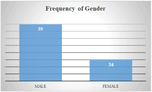
73 children of VSD were selected in the study. 53.4% were male and female were 46.6% (Fig 1).
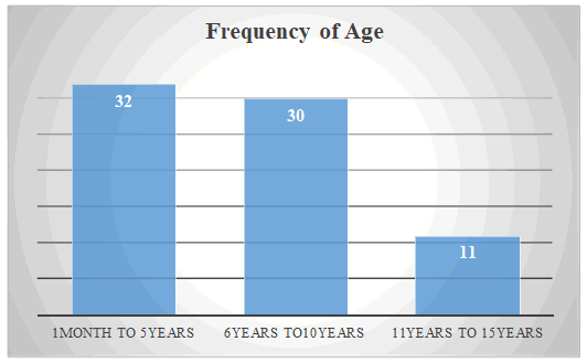
According to age breakdown, 32 (43.8%)children belong to 1 month to 5 years, 30(40%) children were belong to 6 years to 10 years and 11(16%) were from 11years to 15 years (Fig2).
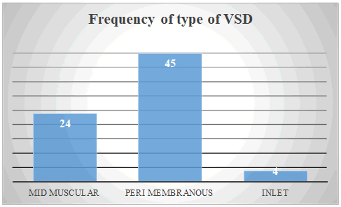
Regarding types of VSD, 61% (45) were peri-membranous type of VSD, 32% (24) were having mid muscular type of VSD, and 5.4% (4) were inlet type of VSD (Fig 3).
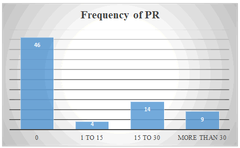
Based on pulmonary regurgitation 5.4% children had mild pulmonary hypertension, 19% had moderate pulmonary hypertension and 12.3% children reflected severe pulmonary hypertension (Fig 4)
The result of echocardiography revealed that LVEDD mean was 39.4 mm with standard deviation of 8.96. Longitudinal Strain had with mean -19.78%, LV mass had mean of 56.75g and LV mass index had mean of 70.43g/m² (Table 1)
Table 1: Descriptive Statistics of Quantitative Variables
| Descriptive Statistics of Quantitative Variables | |||||
|---|---|---|---|---|---|
| N | Minimum | Maximum | Mean | Std. Deviation | |
| VSD size in mm | 73 | 2.00 | 14.80 | 6.5384 | 3.42518 |
| LV end diastolic dimension in mm | 73 | 12.00 | 52.00 | 34.9863 | 8.96830 |
| LV end systolic dimension in mm | 73 | 7.00 | 62.00 | 29.4247 | 12.74637 |
| Ejection fraction in % | 73 | 59.00 | 82.00 | 67.2329 | 6.97719 |
| Mitral annular plane systolic excursion in mm | 73 | 12.00 | 31.00 | 21.5342 | 5.31006 |
| Tricuspid annular plane systolic excursion in mm | 73 | 10.00 | 39.00 | 23.8767 | 5.00817 |
| Longitudinal Strain of LV in % | 73 | -27.00 | -11.30 | -19.7877 | 4.14548 |
| Left ventricle mass in grams | 73 | 8.00 | 168.00 | 56.7534 | 32.07057 |
| Left ventricle mass index of patient in gram/m² | 73 | 19.00 | 150.00 | 70.4384 | 31.37275 |
| Valid N (list wise) | 73 | ||||
LVEDD obtained in this study with mean of 34.98mm having positive correlation with the VSD sizes, which showed that LVEDD increase with increase in size of VSD. The average LV strain was -19.78 which is quite normal but in correlation with VSD size, it showed negative correlation showing LV strain decrease at some extent with increase in VSD sizes (Table 2)
Table 2:Correlation of VSD size with Longitudinal Strain and LVEDD
| Pearson correlation | P value | |
|---|---|---|
| Longitudinal strain of LV | -0.366 | 0.001 |
| LV end diastolic dimension in mm | 0.419 | 0.000 |
DISCUSSION
For CHD it was still uncertain how regional dysfunction affects global function and the role of regional differences during the progression of myocardial dysfunction need to be defined. The new methods also allow quantification of myocardial motion and deformation in different directions (longitudinal, radial, and circumferential) while conventional methods mainly rely on the assessment of radial function [4]. Still, isovolumic acceleration (IVA), derived from the early systolic tissue Doppler velocity tracing, and peak systolic strain rate, both of which normally occur during early systole, were relatively independent of loading conditions [5], [6]
Strain and strain rate revealed sensitive parameter of systolic function [2]. We also found that the patients with VSDs have a greater LVEDD than usual individuals. As far as LV strain concerns the normal value should be up to -20.0% in normal children. In my study, average LV strain is -19.78 which is quite normal but in correlation with VSD size, it showed negative correlation that mean LV strain decreased at some extent with increased in VSD size. Our study favor Jamie Penk et al1 study which showed in their study that there was no change in the average longitudinal strain and strain rate of six segments in the LV. Kimbell TR also assess the contractility and found no difference in patients with VSD versus normal children [7].Same result favored our study which also revealed no significant difference in both longitudinal strain and LVEDD. Corin WJ et al. also found no change in contractility between normal children and those having VSD with congestive heart failure by measuring mean velocity of circumferential shortening [8]. Our study supported these result using longitudinal strain and strain rate.
Kleinman C et al. noticed increased in LVEDD in children with VSD [9] .We also noticed same result in our study. No difference in strain or strain rate shown between the genders and with age as also supported by Lorch S et al. [10]. As far as LV strain concerns its normal value should be up to -20.0% in normal children. Our study showed average LV strain is -19.78 which is quite normal but in correlation with VSD size it shows negative correlation showing LV strain decrease at some extent with increase in VSD size.
CONCLUSION
This recommends that contractility assessed by echocardiography did not change by a hemodynamically large VSD when assessed by longitudinal strain and stain rate.
References
- Jamie Penk, Angira Patel, Amy Lay, Catherine Webb (2014) Longitudinal Strain and Strain Rate in patients with Hemodynamically significant Ventricular Septal Defect:World. Journal for Pediatric and Congenital Surgery. 5:2 216-218
- Geyer H, Caracciolo G, Abe H, et al. (2010) Assessment of myocardialmechanics using speckle-tracking echocardiography: fundamentals and clinical applications. J Am Soc Echocardiogr. 23:4 351-369
- Missant C, Rex S, Claus P, Mertens L, Wouters PF et al. (2008) Load-sensitivity of regional tissue deformation in the right ventricle: isovolumic versus ejection-phase indices of contractility. Heart (British Cardiac Society) 94:15
- Mark K. Friedberg and Luc Mertens (2009) Tissue velocities, strain, and strain rate for echocardiographic assessment of ventricular function in congenital heart disease. European Journal of Echocardiography. 10:5 85â??93
- Lee TY, Kang PL, Hsiao SH, Lin SK, Mar GY, Chiou CW et al. (2007) Tissue Doppler velocity is not totally preload-independent: a study in a uremic population after hemodialysis. Cardiology. 107:415â??421
- Becker M, Kramann R, Dohmen G, Luckhoff A, Autschbach R, Kelm M et al. (2007) Impact of left ventricular loading conditions on myocardial deformation parameters: analysis of early and late changes of myocardial deformation parameters after aortic valve replacement. J Am Soc Echocardiogr. 20:6 681â??689
- Kimball TR, Daniels SR, Meyer RA, Hannon DW, Khoury P,Schwartz DC et al. (1991) Relation of symptoms to contractility and defect size in infants with ventricular septal defect. Am J Cardiol. 67:13 1097-1102
- Corin WJ, Swindle MM, Spann JF, et al. (1988) Mechanism of decreasedforward stroke volume in children and swine with ventricular septal defect and failure to thrive. J Clin Invest. 82:2 544-551
- Kleinman C, Tabibian M, Starc T, Hsu D, Gersony W et al. (2007) Spontaneous regression of left ventricular dilation in children with restrictive ventricular septal defects. J Pediatr. 150:6 583-586
- Lorch S, Ludomirsky A, Singh G (2008) Maturational and growth-relatedchanges in left ventricular longitudinal strain and strain rate measured by two-dimensional speckle tracking echocardiography in healthy pediatric population. J Am Soc Echocardiogr. 21:11 1207-1215
Open Access Journals
- Aquaculture & Veterinary Science
- Chemistry & Chemical Sciences
- Clinical Sciences
- Engineering
- General Science
- Genetics & Molecular Biology
- Health Care & Nursing
- Immunology & Microbiology
- Materials Science
- Mathematics & Physics
- Medical Sciences
- Neurology & Psychiatry
- Oncology & Cancer Science
- Pharmaceutical Sciences


