A rare case of right ventricular myxoma in a child
Ayad Anass
Cardiovascular surgery unit, Military hospital of Rabat, Morocco
- *Corresponding Author:
- Ayad Anass
Unit, Military hospital of Rabat, Morocco
E-mail: drayadanass@gmail.com
Received: September 14, 2020; Accepted: September 15, 2020; Published: September 30, 2020
Citation: Anass A (2020) A rare case of right ventricular myxoma in a child. Vol.S No.1: 1 DOI: 10.36648/IPIPC.S.1.004
Copyright: ©2020 Anass A . This is an open-access article distributed under the terms of the Creative Commons Attribution License, which permits unrestricted use, distribution, and reproduction in any medium, provided the original author and source are credited.
Abstract
Myxoma of the right ventricle is quite rare and have not been reported in children. The most common complications are pulmonary embolism and obstruction the pulmonary valve. We report the case of a 06-year-old child admitted to Cardiology for palor and asthenia without any episode of syncope. Transthoracic echocardiography revealed a mass of 2cm arising from the tricuspid valve and projecting through the infundibulum . Surgical excision was performed under extracorporeal circulation,. pathological studies made it possible to confirm the diagnosis of myxoma.
Introduction
Myxomas are the most common cardiac tumors in adults, but are rare during infancy and childhood. The prevalence of cardiac myxomas varies between 0.0017% and 0.19% in autopsy studies [1]. Only 5% of these tumors are found in the right ventricle (RV) [2]. Cardiac tumors originating from the right ventricular outflow tract (RVOT) present unusual diagnostic and therapeutic challenges. The symptoms are dependent on the size and location of the tumor. Such tumors are capable of generating major clinical consequences, including arrhythmias, pulmonary emboli, and sudden death. Once the diagnosis of the tumor obstructing the RVOT is confirmed, immediate surgery is indicated. Here we report a case of a primary cardiac myxoma located in the RVOT
Case presentation
An 11-year-old boy was referred to our department for the exploration of syncope. There was no family history of myxoma. On admission, his pulse rate was 100 beats/minute and blood pressure was 100/60mmHg. He had no signs of congestive cardiac failure. On physical examination, a grade 4/6 systolic ejection murmur was heard at the left upper sternal border. His chest radiography was normal. His electrocardiogram showed normal sinus rhythm of 110 beats/minute and ST segment depression in leads V1-V4 with right ventricular hypertrophy and right axis deviation.
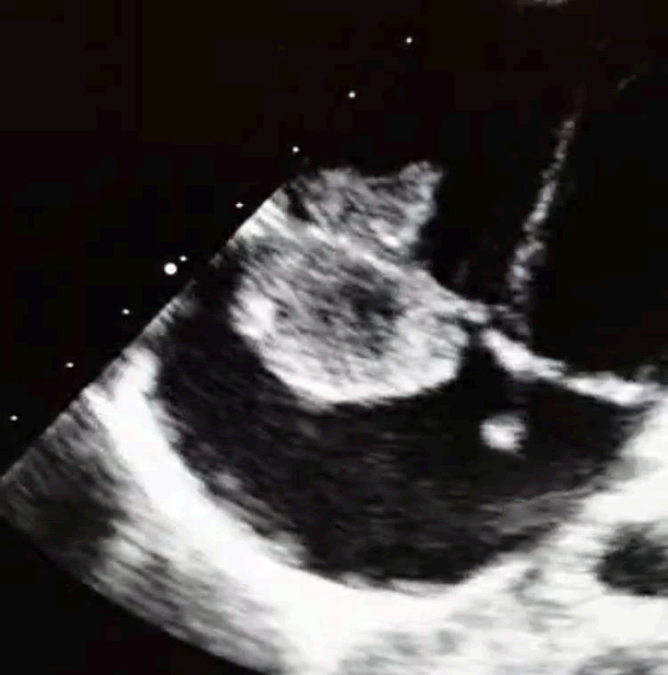
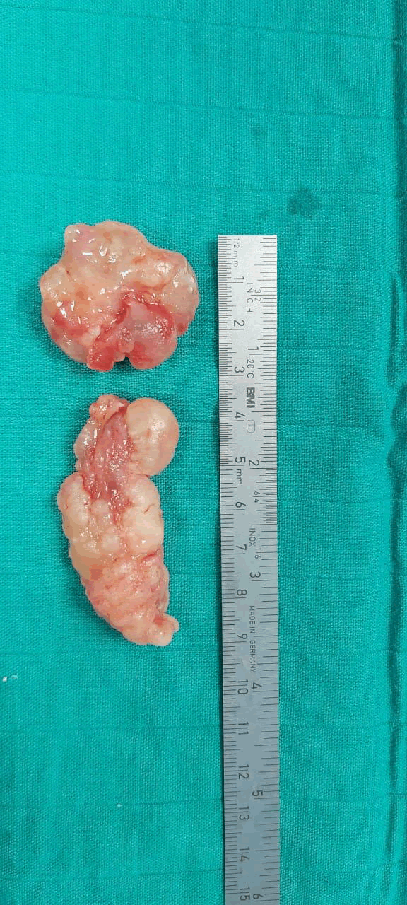
Transthoracic echocardiography revealed a large mobile 19×19mm mass attached to RVOT. The pulmonary valve seemed to be spared. The tumor was causing an obstruction of the RVOT (Figure 1). His RV-to-pulmonary artery pressure gradient was 90mmHg. His echocardiography showed right atrium (RA) and RV dilatation, mild tricuspid regurgitation with right ventricular systolic pressure at 105mmHg. Abdominal ultrasound and venous Doppler of both his lower limbs were normal. He was anaesthetized, perioperative cardiac arrest occurred, emergency sternotomy and cardiopulmonary bypass (CPB) was established through aortic and bicaval cannulation. His RA was opened. Operative findings revealed a gelatinous 20×20mm mass, originating 1cm below the pulmonary valve and attached to the RVOT on its septal surface (Figure 2). The mass was completely removed. The diagnosis of myxoma was confirmed by histology. The postoperative period was uneventful and he was discharged after 7 postoperative days. Postoperative echocardiography documented an unobstructed RVOT.
Discussion
Primary cardiac masses are rare and usually benign with myxoma being the most common [1]. Up to 75% of myxomas are located in the left atrium whereas right-sided myxomas are rare (15 to 20%), with right ventricular (3 to 4%) or pulmonary artery myxomas being extremely rare [3]. There are several case reports of right ventricular myxoma obstructing the outflow tract in adults and older children, but reports of this pathology during early childhood are scarce; cardiac myxomas in the pediatric age group have relevance in particular to a condition known as familial myxoma syndrome. This syndrome, also known as Carney’s syndrome, consists of a variable complex of mucocutaneous, visceral, and endocrine disorders. Symptoms may be variable and are determined by the tumor location and size. Right ventricular myxomas may cause obstruction of the RVOT and pulmonary main trunk, which may also lead to complications such as syncope, pulmonary embolism, and sudden death. Cardiac myxoma extending into the RVOT is a rare cause of right heart failure. Symptoms and signs include peripheral edema, ascites, and shortness of breath as a result of RVOT obstruction. The gold standard noninvasive diagnostic modality for such tumors is transthoracic or transesophageal echocardiogram. An echocardiogram enables preoperative localization of the tumor, size, shape, mobility as well as the risk of RVOT obstruction and the tumor attachment, and therefore facilitates the selection of the optimal surgical approach. Transesophageal echocardiography accurately identifies other localization of myxomas. Cardiac-gated computed tomography and cardiac-gated magnetic resonance scans offer additional information about the structure and function of cardiac tumors before surgical resection [4]. The differential diagnosis for an intracavitary cardiac mass includes thrombus, myxoma, lipoma and nonmyxomatous neoplasm, most of which are malignant [5]. The risk of life-threatening complications indicates the importance of early diagnosis and prompt surgical resection as soon as possible. One of the main concerns during anesthesia is the risk of tumor embolization and pulmonary obstruction during anesthesia induction or at any stage thereafter. Hemodynamic instability, inconvenient manipulation of the tumor and heart must be avoided to prevent these complications. Femoral arterial-venous cannulation can be performed for CPB initiation in the case of complete RVOT obstruction with hemodynamic collapse. Several surgical techniques have been suggested, but in each case it depends on the site of the tumor. Follow up for recurrent myxoma in an uncommon location is recommended.
Conclusions
Childhood cases of right ventricular myxoma obstructing the RVOT are rare. Immediate, careful surgical resection should be performed to avoid outflow tract obstruction, which might otherwise result in sudden death.
References
- Gopal AS, Stathopoulos JA, Arora N, Banerjee S, Messineo F. Differential diagnosis of intracavitary tumors obstructing the right ventricular outflow tract. J Am Soc Echocardiogr. 2001;14(9):937–940. doi: 10.1067/mje.2001.114627. [PubMed] [CrossRef] [Google Scholar]
- Hirota J, Akiyama K, Taniyasu N, Maisawa K, Kobayashi Y, Sakamoto N, Komatsu N. Injury to the tricuspid valve and membranous atrioventricular septum caused by huge calcified right ventricular myxoma: report of a case. Circ J. 2004;68(8):799–801. doi: 10.1253/circj.68.799. [PubMed] [CrossRef] [Google Scholar]
- Huang SC, Lee ML, Chen SJ, Wu MZ, Chang CI. Pulmonary artery myxoma as a rare cause of dyspnea for a young female patient. J Thorac Cardiovasc Surg. 2006;131(5):1179–1180. doi: 10.1016/j.jtcvs.2005.12.052. [PubMed] [CrossRef] [Google Scholar]
- Lacey BW, Lin A. Radiologic evaluation of right ventricular outflow tract myxomas. Tex Heart Inst J. 2013;40(1):68–70. [PMC free article] [PubMed] [Google Scholar]
- GElderkin RA, Radford DJ. Primary cardiac tumours in a paediatric population. J Paediatr Child Health. 2002;38(2):173–177. doi: 10.1046/j.1440-1754.2002.00734.x. [PubMed] [CrossRef] [Google Scholar]
Open Access Journals
- Aquaculture & Veterinary Science
- Chemistry & Chemical Sciences
- Clinical Sciences
- Engineering
- General Science
- Genetics & Molecular Biology
- Health Care & Nursing
- Immunology & Microbiology
- Materials Science
- Mathematics & Physics
- Medical Sciences
- Neurology & Psychiatry
- Oncology & Cancer Science
- Pharmaceutical Sciences


